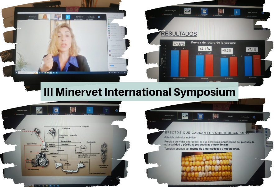
III Minervet International Symposium
28 de June de 2022
First session of the III Minervet International Symposium
22 de July de 2022A necropsy is the post-mortem examination of an animal to determine the cause of disease or death. It involves the regulated dissection of the animal and the performance of a detailed macroscopic examination of all organ systems. The tissue samples taken during the necropsy are sent to the Pathological Anatomy laboratory to carry out the histopathological study and/or to other laboratories to carry out complementary diagnostic tests (microbiology, toxicology, etc.) When a necropsy is carried out, it is intended to answer the question of what has been the cause of death in the animal.
The necropsy allows us to know, in some cases, if the clinical diagnosis is correct. In daily practice, knowing the cause of death of an animal makes it possible to establish therapeutic or preventive measures for the group from which it comes. Likewise, necropsy can be a useful control measure to evaluate the different established treatments. The necropsy allows us to advance in the knowledge of the disease, in relation to the pathogenesis of the lesions produced, and their association with specific etiological agents.
In veterinary medicine it is common for professionals to perform necropsies frequently. Hence the interest in unifying value criteria in pathological diagnosis and progressing in the knowledge of diseases through the macroscopic lesions that can be observed.
Performing a necropsy does not entail major complications in general; however, if valid conclusions are to be drawn, one needs to proceed in a certain order and according to a method. In fact, there are different procedures to perform a necropsy, although they all share common factors:
• Systematic necropsy; it is necessary to use a system, and apply it in the same way in all cases.
• Regulated necropsy; it is necessary to follow an order in its realization.
• Complete necropsy; Avoid leaving parts or organs of the animal unexamined.
The general scheme of a necropsy must include the following steps:
1. Preparation and external examination of the corpse.
2. Opening the corpse.
3. Study of the organs of the abdominal cavity.
4. Study of the organs of the thoracic cavity.
5. Study of the head.
6. Study of lymph nodes and bone marrow.
7. Study of the locomotor system (bones, joints and muscles).
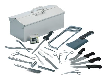
The equipment to perform a proper necropsy is: Gloves, overalls, chinstrap, goggles, cap, face mask, apron, rubber boots, dissection knife and/or scalpel, dissection material, material for taking samples and finally if It is possible an assistant person who will take step-by-step photographs or video filming.

The general rules to follow in a regulated necropsy are:
- Animals that are at the end of an infectious process should not be selected.
- Do not select chronically ill animals
- It is advisable to sacrifice animals in the different stages of the disease, to correlate the clinical symptoms with the lesions that we observe and establish the possible correlation between what is observed in the necropsy and what we see in the farm.
- Do not take samples to carry out antibiograms of treated animals
The general procedure for the preparation and external examination of the corpse is:
- Opening of the corpse.
- Study of the organs of the abdominal cavity.
- Study of the organs of the thoracic cavity.
- Study of the head.
- Study of lymph nodes and bone marrow.
- Study of the locomotor system, bones, joints and muscles.
The external examination of the corpse allows assessing the state of the animal’s meat and the existence of skin lesions: Mucous membranes, nostrils and oral cavity Perineal region, Abdominal and inguinal region, Joints
The positioning of the animal may vary. The supine or lateral decubitus position is generally used. (Intagri) The opening of the cavities will depend directly on the position in which the necropsy is performed. In supine animals, two cuts are initially made on the skin and subcutaneous tissue in the shape of a triangle following the projection of the mandible. Subsequently, an incision is made to the entrance of the thorax, where with the same cutting instrument, the cartilaginous area of the ribs is sectioned, revealing the entire rib cage. Following the cut, the abdominal cavity is also opened up to the pubis.
The correct choice of animals and the performance of a necropsy in a regulated manner, together with the proper collection of samples and their shipment to the laboratory, will provide important information about the pathology that is being suffered.
A sufficient number is necessary (since a small number skew the diagnosis). This number of animals varies, depending on the evolution of the process, whether it is an overacute process or a slow evolution process on the farm (Toledo Castillo et al. 2015).
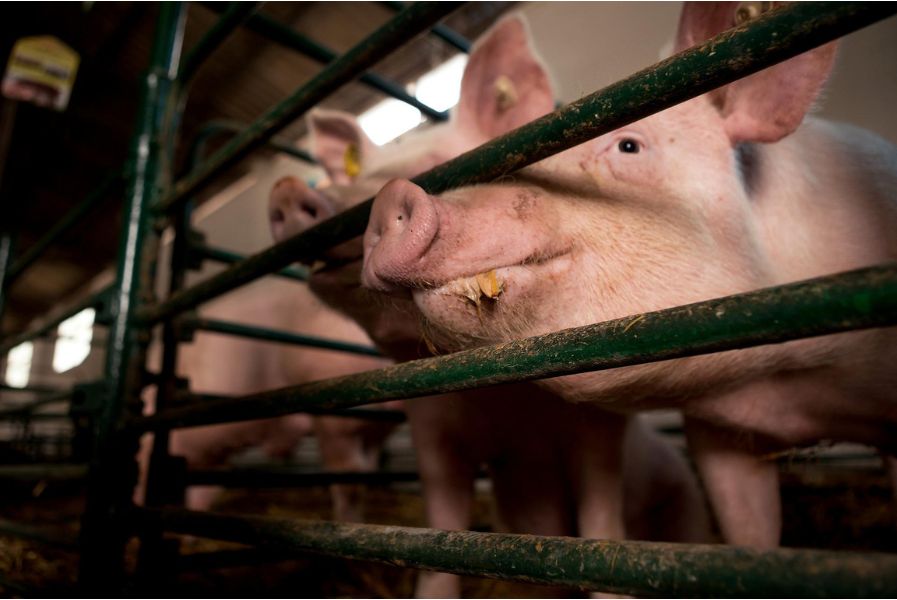
Initially, it is necessary to verify the degree and extension of the cadaveric changes, which will give approximate information on the time elapsed since the death of the animal and, above all, on the state of decomposition of the corpse. Corpses in an advanced stage of autolysis do not usually offer much information and can lead to confusion when the macroscopic diagnosis has to be established (it is difficult to determine if these are changes due to the disease or the autolysis process). The sample considered ideal is the live pig, because the clinical symptoms it presents can be observed (it gives an idea of whether it is a representative animal of the farm’s problem or not). In addition, it is possible to draw blood to determine some serum or blood parameter.
For the necropsy, the animal would be euthanized and immediately bled. Both animals for slaughter and those used for experimentation or other scientific purposes must be slaughtered using a method that does not cause pain or suffering.
Taking into account all of the above, the following slaughter methods are especially discouraged:
• Administration of euthanasia drugs for veterinary use.
• Live bleeding (bleeding may be acceptable if the animal has been previously stunned).
• Head trauma. Post-mortem changes can give us an idea of how long an animal has been dead. Among the post-mortem changes that must be observed, the following stand out:
• Cadaveric rigidity (rigor mortis).
• Body temperature.
• Corneal opacity and loss of eye turgidity
• Presence and magnitude of signs of decomposition (green abdominal spots). Next, a complete external examination (skin, hair, superficial lymph nodes) is performed from the skull to the caudal region.
Sending samples to the laboratory
• It is necessary to call the laboratory and ask what samples they need to identify the process and their shipping conditions.
• Secondly, having the material ready: sterile bags or jars (to send pieces, fluids or organs) and formaldehyde in the trunk of the car is always useful.
It is advisable to perform antibiograms for some infectious processes so that antibiotic treatments adjusted to sensitivity tests can be established in the future. (Toledo, Manuel et al.2015) Histopathology is an excellent diagnostic tool to guide pathological problems of difficult clinical characterization and problems of neurological origin. Logically, it also allows the correlation between the detection of infectious agents and the existence of associated injuries.
Bibliography
Intagri Técnica de necropsia en Cerdos PorciNews.Porcino.Infode https://www.intagri.com/articulos/ganaderia/tecnica-de-necropsia-en-cerdos –
Toledo Castillo,Manuel et al. Necropcia de campo en ganado porcino(2015)Revista porciNews https://porcino.info/necropsia-de-campo-en-ganado-porcino
Author: MV. Germán González

